AI-Powered LC-OCT Skin Image Analysis for Research
Turn LC-OCT images into research-grade quantitative data.
SkinArch™ pairs advanced LC-OCT imaging with AI-assisted segmentation and quantitative analysis, so researchers can visualise skin microstructure, evaluate treatment efficacy, and produce reproducible data — without a biopsy.
For Research Use. Not for diagnostic or therapeutic use.
Why SkinArch™?
AI-Powered Skin Image Analysis
One platform for LC-OCT image analysis — import, segment, reconstruct, measure, visualize, and report.
Image Import & Standardization
Prepare LC-OCT stacks so every study runs on the same footing.
AI Skin Layer Segmentation
Key structures identified automatically, reviewable frame by frame.
3D DEJ Reconstruction
The dermal-epidermal junction rebuilt as an interactive 3D surface.
Quantitative Analysis
Thickness, surface morphology and structural metrics, measured consistently.
2D & 3D Visualization
Move between cross-section, en-face and volume views of the same scan.
Research Report Export
Standardised outputs your study documentation can cite directly.
The platform
Streamlined LC-OCT Analysis Workflow
From image acquisition to quantitative analysis, SkinArch™ simplifies the skin imaging research workflow.
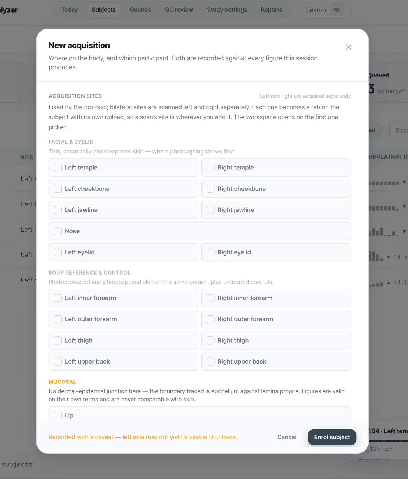
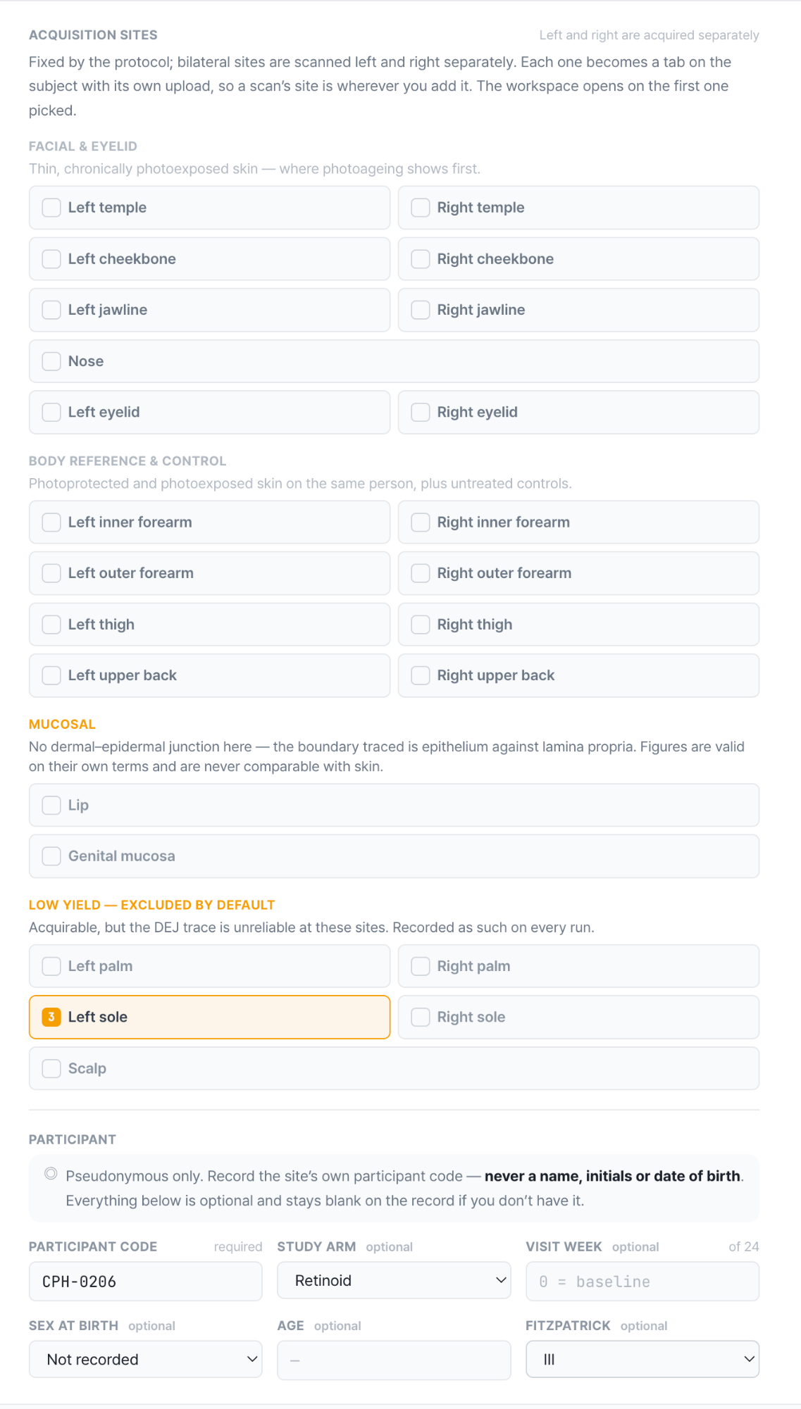
Enroll (new acquisition)
Pick the acquisition sites and record the participant code. Sites are fixed by the protocol, and every figure the session produces is filed against both.
Run analysis
Add .tif volumes to the stack and press once. The junction is traced slice by slice while you watch progress and the queue in the same view.
227 queued · DEJ-Net v2.4
Applications & benefits
LC-OCT Applications in Cosmetics & Skin Research
From early formulation screening to clinical validation, SkinArch™ turns LC-OCT images into objective, comparable data.
Anti-aging studies
Monitor structural skin changes over time.
Hydration research
Evaluate skin response to moisturisation treatments.
Barrier function assessment
Analyse skin integrity and recovery.
Ingredient efficacy
Quantify the effects of cosmetic formulations.
Clinical product validation
Generate objective data to support research claims.
Longitudinal studies
Compare the same skin region throughout treatment.
Why LC-OCT?
High-Resolution LC-OCT Skin Imaging Without Biopsy
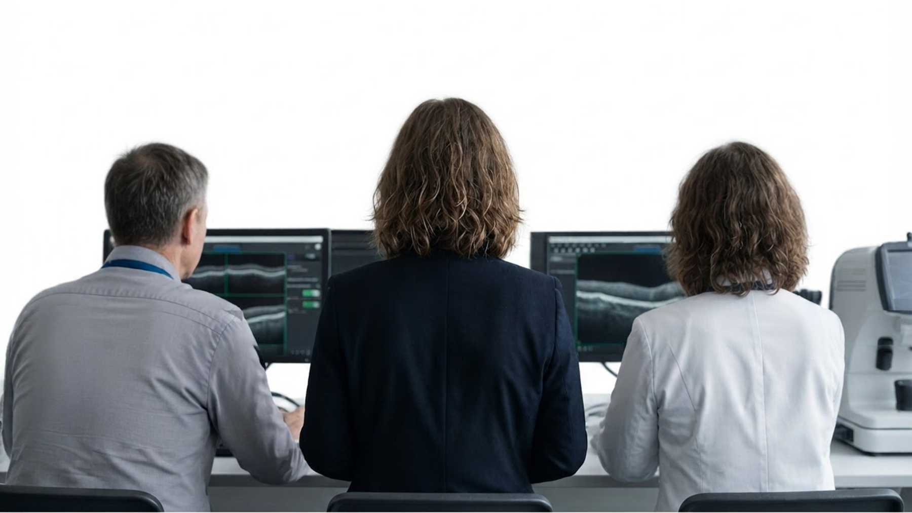
LC-OCT images living skin at high resolution, so microscopic structures can be evaluated in real time with no tissue removed.
Research workflow
LC-OCT scan
Capture a vertical section of living skin at the study site.
AI segmentation
Layers and the dermal-epidermal junction are traced automatically.
3D reconstruction
The junction is rebuilt as a surface you can rotate and inspect.
Quantitative analysis
Thickness, morphology and structural metrics are computed per scan.
Visualisation
Cross-section, en-face and volume views of the same dataset.
Research report
Standardised output ready for study documentation.
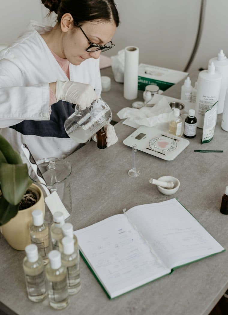
Cosmetics R&D
Evaluate ingredients and formulations using quantitative skin structure data.
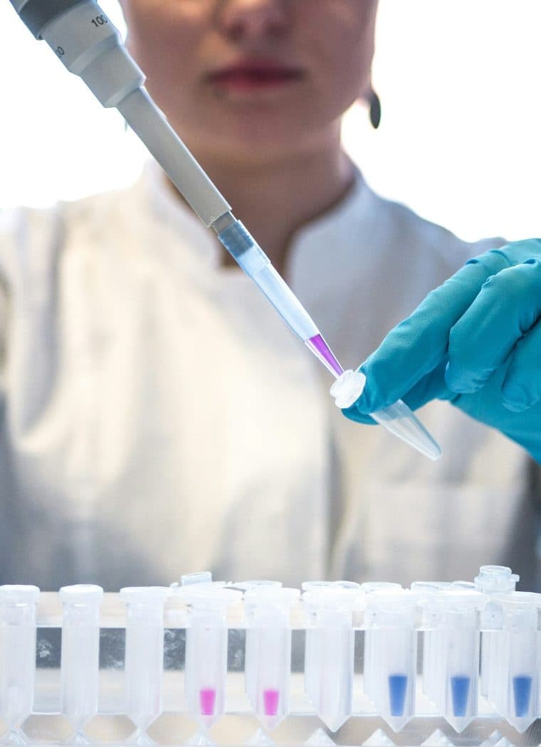
Clinical Research
Support product validation with objective, comparable research outputs.
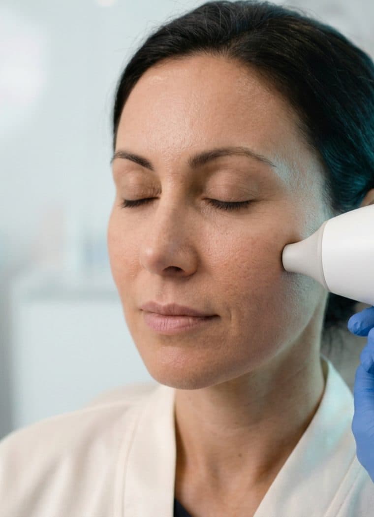
Skin Science Teams
Visualise and analyse skin microstructure across research timepoints.
Built for research teams
Skin Imaging Software for Research Teams
SkinArch™ can support teams working across cosmetic formulation research, product validation, and skin science.
FAQ
Frequently Asked Questions
What research teams most often ask before their first LC-OCT analysis.
What is LC-OCT skin imaging?
Line-field confocal optical coherence tomography (LC-OCT) is a non-invasive imaging method that captures skin structure at high resolution without a biopsy. It produces stacks of cross-sectional images through the skin's layers.
What does SkinArch measure from LC-OCT images?
SkinArch identifies skin layers — surface, stratum corneum and the dermal–epidermal junction (DEJ) — and produces quantitative outputs including undulation, thickness and surface area, alongside 2D overlays and 3D visualizations.
How is this different from reviewing LC-OCT images manually?
Manual slice-by-slice tracing is slow and varies between reviewers. SkinArch applies the same automated analysis across a whole stack, so results stay consistent between runs and between reviewers.
Can SkinArch be used in clinical studies?
SkinArch is intended for research use only and is not intended for diagnostic or therapeutic use. Research teams use it as an additional objective measurement layer alongside their existing assessments.
Does W Labs supply the LC-OCT imaging device?
No. SkinArch is analysis software. It works with LC-OCT images your team has already acquired.
Who is SkinArch built for?
Skin research teams in cosmetics R&D, dermatology and clinical research, and imaging or analysis groups working with LC-OCT data.

Explore AI-Powered LC-OCT Skin Analysis
See how SkinArch™ can support cosmetics R&D, clinical research, and skin studies with quantitative skin imaging. Request a live demo to see it on your own workflow.